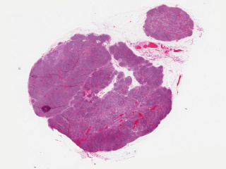
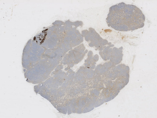
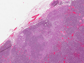
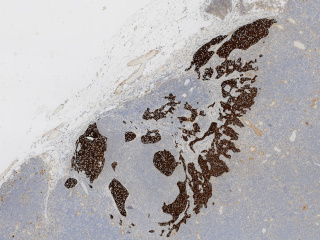
Whole slide imaging of 396 full cases of axillary lymph nodes in breast cancer cases. Included are both sentinel node surgery and axillary dissections pre, peri or post breast cancer surgery or treatment. Sentinel node cases are cut in three levels (stained with HE) and one additional slide immunohistochemically stained with CKAE1/AE3. The number of sentinel node cases with complete immunohistochemical staining is 321. The axillary dissections are cut with one cut level as default. No frozen sections included. The cases are anonymised and exported from the digital archive at the Department of Clinical Pathology in Linköping, Region Östergötland. Included are both positive and negative cases. Some metadata on case level is available (positive or negative case, number of nodes, primary tumour and if neoadjuvant treatment in axillary dissections was given).
Keywords: Pathology, Whole slide imaging, Breast, Lymph nodes, Cancer, Sentinel nodes, Immunohistochemical staining, cytokeratin, CKAE1/AE3.
Sample images
Sample images with reduced image quality. Please click to preview.
Dataset information
| Short name | BRLN |
|---|---|
| Origin | Clinical |
| Cite as |
Sofia Jarkman, Martin Lindvall, Joel Hedlund, Darren Treanor, Claes Lundstrom, and Jeroen van der Laak
(2019)
Axillary lymph nodes in breast cancer cases
doi:10.23698/aida/brln [BibTeX format] |
| Field | Pathology |
| Organ |
Breast |
| Age span | - |
| Title | Axillary lymph nodes in breast cancer cases |
| Author |
Sofia Jarkman
Martin Lindvall Joel Hedlund Darren Treanor Claes Lundstrom Jeroen van der Laak |
| Year | 2019 |
| DOI | doi:10.23698/aida/brln |
| Status | Ongoing |
| Version | 1.0.2 |
| Scans | 4462 |
| Annotations | 0 |
| Size | 2.36TB |
| Resolution | 20x |
| Modality |
SM
|
| Scanner |
Aperio ScanScope AT Hamamatsu NanoZoomer XR Hamamatsu NanoZoomer S360 Hamamatsu NanoZoomer S60 |
| Stain | Hematoxylin and eosin. In sentinel node cases also immunohistochemical stain for cytokeratin AE1/AE3. |
| Phase | |
| References |
|
| Copyright | Copyright 2019 Linköping University, Claes Lundström |
| Access |
Available under the following licenses, described in the License section below.
Controlled access
AIDA BY CA license
|
Annotation
No in-image annotations available.
File formats
Pixel position scaling
Coordinates given are relative to the image width. To get the correct pixel position, X coordinates (and Y coordinates!) should therefore be multiplied with the image width.
License
Controlled access
Free for use in legal and ethical medical diagnostics research.
To request access to the dataset, use the Apply for access button below.
Note that the recipient researcher must hold at least a PhD degree in a
relevant field and that the applicant should be an authorized signatory who can
legally enter into data sharing agreements on the behalf of the institution.
Help with applying for controlled access.
AIDA BY CA license
Copyright 2019 Linköping University, Claes Lundström
Permission to use, copy, modify, and/or distribute this data within Analytic Imaging Diagnostics Arena (AIDA) for the purpose of medical diagnostics research with or without fee is hereby granted, provided that the above copyright notice and this permission notice appear in all copies, and that publications resulting from the use of this data cite the following works:
Sofia Jarkman, Martin Lindvall, Joel Hedlund, Darren Treanor, Claes Lundstrom, and Jeroen van der Laak (2019) Axillary lymph nodes in breast cancer cases doi:10.23698/aida/brln.
THE DATA IS PROVIDED “AS IS” AND THE AUTHOR DISCLAIMS ALL WARRANTIES WITH REGARD TO THIS DATA INCLUDING ALL IMPLIED WARRANTIES OF MERCHANTABILITY AND FITNESS. IN NO EVENT SHALL THE AUTHOR BE LIABLE FOR ANY SPECIAL, DIRECT, INDIRECT, OR CONSEQUENTIAL DAMAGES OR ANY DAMAGES WHATSOEVER RESULTING FROM LOSS OF USE, DATA OR PROFITS, WHETHER IN AN ACTION OF CONTRACT, NEGLIGENCE OR OTHER TORTIOUS ACTION, ARISING OUT OF OR IN CONNECTION WITH THE USE OR CHARACTERISTICS OF THIS DATA.