This dataset consists of 361 whole slide images (WSI) - 296 malignant from women with invasive breast cancer (HER2 neg) and 65 benign. The tumours have been classified with four SNOMED-CT categories based on morphology: invasive duct carcinoma, invasive lobular carcinoma, in situ carcinoma, and others. 4144 separate annotations have been made to segment different tissue structures connected to ontologies.
Keywords: Pathology, Breast, Cancer, Whole slide imaging, Annotated.
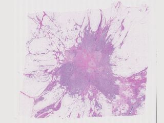
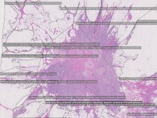
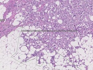
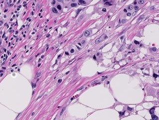
Sample images
Sample images with reduced image quality. Please click to preview.
Dataset information
| Short name | DRBR |
|---|---|
| Origin | Clinical |
| Cite as |
Anna Bodén, Jerónimo F. Rose, Martin Lindvall, and Caroline Bivik Stadler
(2019)
Breast data from the Visual Sweden project DROID
doi:10.23698/aida/drbr [BibTeX format] |
| Field | Pathology |
| Organ |
Breast |
| Age span | |
| Title | Breast data from the Visual Sweden project DROID |
| Author |
Anna Bodén
Jerónimo F. Rose Martin Lindvall Caroline Bivik Stadler |
| Year | 2019 |
| DOI | doi:10.23698/aida/drbr |
| Status | Completed |
| Version | 1.1.0 |
| Scans | 361 |
| Annotations | 4144 |
| Size | 500.8GB |
| Resolution | 40X single plane, scanned in NDP format |
| Modality |
SM
|
| Scanner |
Hamamatsu NanoZoomer-XR C12000 series 2013 Hamamatsu NanoZoomer 2.0 HT C9600 series 2013 |
| Stain | H&E (hematoxylin and eosin) |
| Phase | |
| References | |
| Copyright | Copyright 2019 Linköping University, Claes Lundström |
| Access |
Available under the following licenses, described in the License section below.
Controlled access
AIDA BY license
|
Annotation
The breast tumours were classified with four SNOMED-CT categories based on morphology: invasive duct carcinoma, Invasive lobular carcinoma, in situ carcinoma and others. 4144 separate annotations were made to segment different tissue structures connected to ontologies. One physician were responsible for the manual annotations controlled by a second pathologist.
File formats
Pixel position scaling
Coordinates given are relative to the image width. To get the correct pixel position, X coordinates (and Y coordinates!) should therefore be multiplied with the image width.
License
Controlled access
Free for use in legal and ethical medical diagnostics research.
To request access to the dataset, use the Apply for access button below.
Note that the recipient researcher must hold at least a PhD degree in a
relevant field and that the applicant should be an authorized signatory who can
legally enter into data sharing agreements on the behalf of the institution.
Help with applying for controlled access.
AIDA BY license
Copyright 2019 Linköping University, Claes Lundström
Permission to use, copy, modify, and/or distribute this data within Analytic Imaging Diagnostics Arena (AIDA) for the purpose of medical diagnostics research with or without fee is hereby granted, provided that the above copyright notice and this permission notice appear in all copies, and that publications resulting from the use of this data cite the following works:
Anna Bodén, Jerónimo F. Rose, Martin Lindvall, and Caroline Bivik Stadler (2019) Breast data from the Visual Sweden project DROID doi:10.23698/aida/drbr.
THE DATA IS PROVIDED “AS IS” AND THE AUTHOR DISCLAIMS ALL WARRANTIES WITH REGARD TO THIS DATA INCLUDING ALL IMPLIED WARRANTIES OF MERCHANTABILITY AND FITNESS. IN NO EVENT SHALL THE AUTHOR BE LIABLE FOR ANY SPECIAL, DIRECT, INDIRECT, OR CONSEQUENTIAL DAMAGES OR ANY DAMAGES WHATSOEVER RESULTING FROM LOSS OF USE, DATA OR PROFITS, WHETHER IN AN ACTION OF CONTRACT, NEGLIGENCE OR OTHER TORTIOUS ACTION, ARISING OUT OF OR IN CONNECTION WITH THE USE OR CHARACTERISTICS OF THIS DATA.