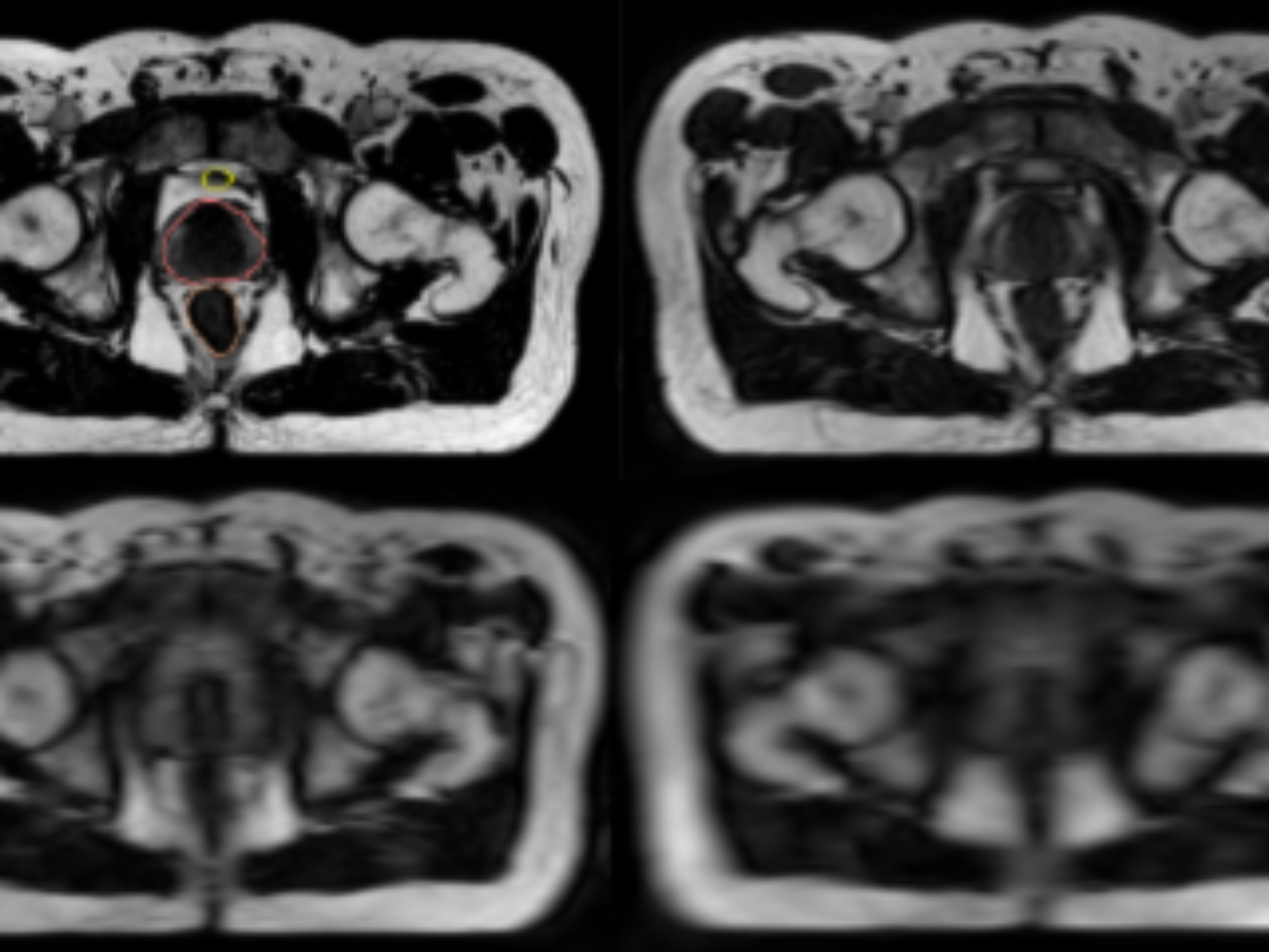
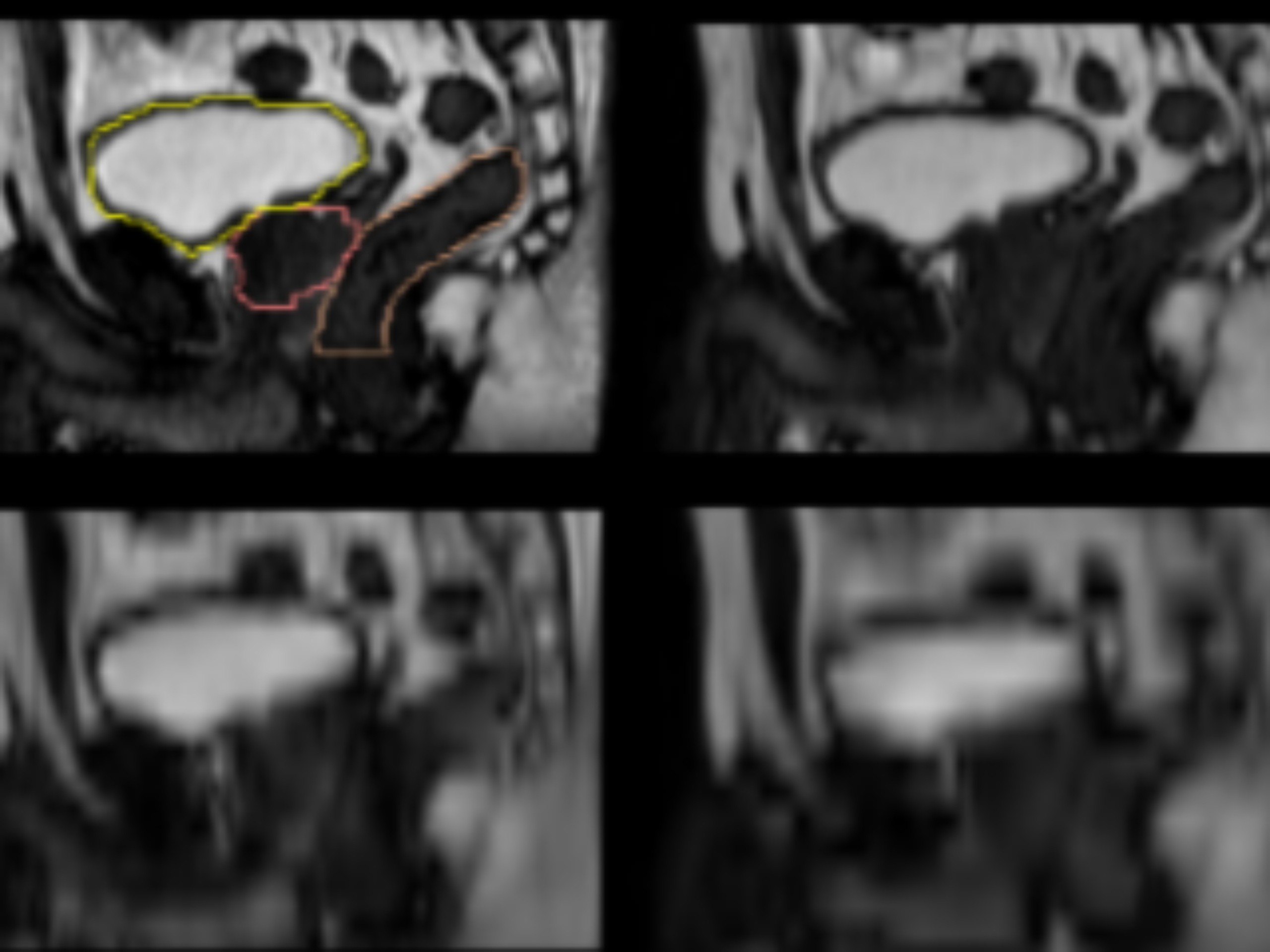
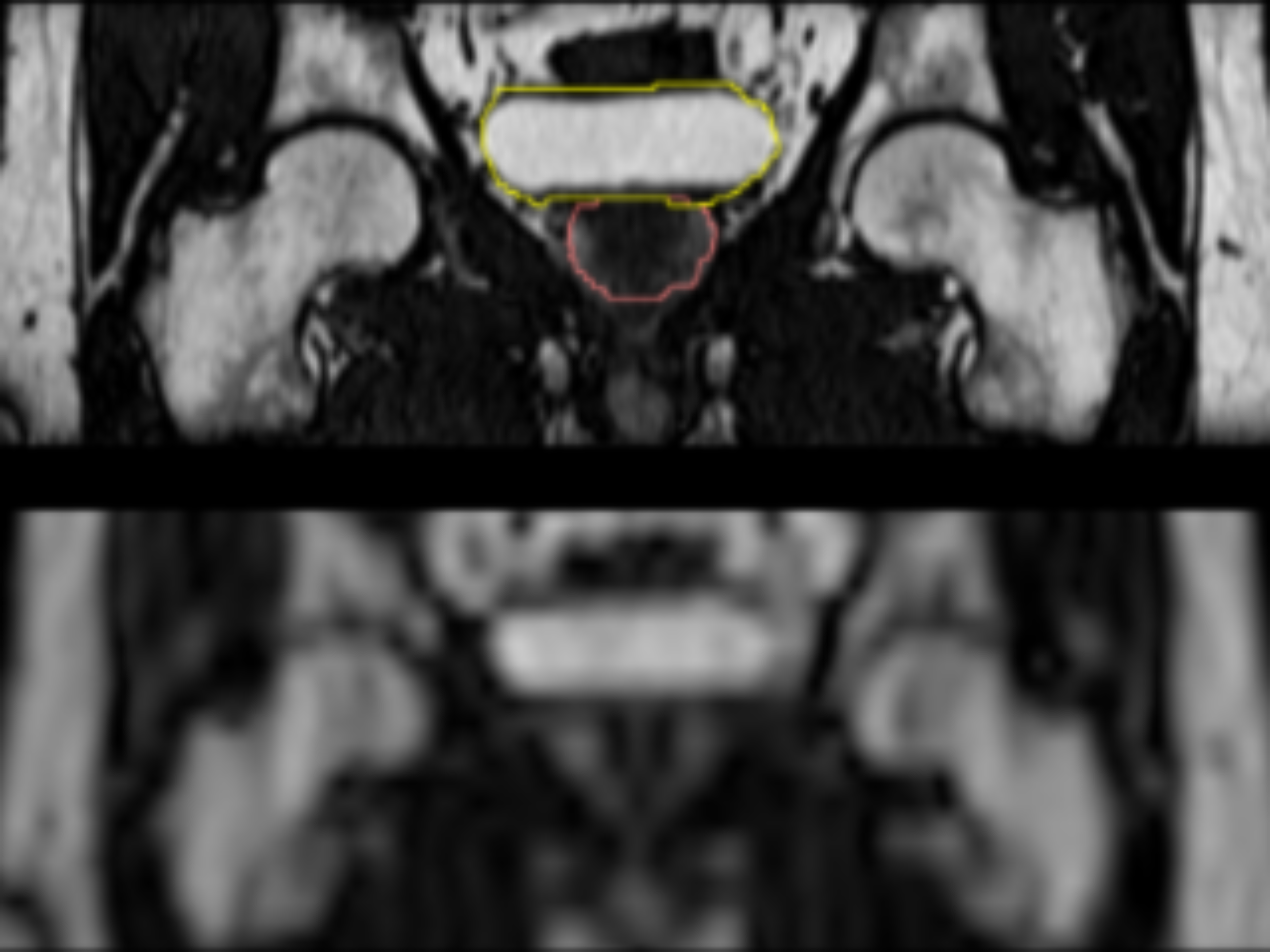
MR-images of the prostate region from healthy volunteers acquired at Elekta unity MR-Linac at Uppsala University Hospital. Data from each volunteer consist of an initial T2-weighted scan, followed by a number of groups of paired low and high resolution data approximately 5 minutes apart with a 3D balanced steady state free precession sequence. The initial T2-image and all high resolution images are segmented by a single observer including prostate, bladder and rectum.
Keywords: Radiology, Annotated, MR-Linac, Radiotherapy, Prostate, Low-resolution.
Sample images
Sample images with reduced image quality. Please click to preview.
Dataset information
| Short name | LESSER |
|---|---|
| Origin | Clinical |
| Cite as |
Samuel Fransson and Robin Strand
(2023)
Low-resolution prostate MR
doi:10.23698/aida/lesser [BibTeX format] |
| Field | Radiology |
| Organ |
Prostate |
| Age span | |
| Title | Low-resolution prostate MR |
| Author |
Samuel Fransson
Robin Strand |
| Year | 2023 |
| DOI | doi:10.23698/aida/lesser |
| Status | Completed |
| Version | 1.0 |
| Scans | 310 |
| Annotations | 255 |
| Size | 5.69GB |
| Resolution | Varying |
| Modality |
MR
|
| Scanner |
Marlin 1.5 T, Elekta Unity |
| Stain | |
| Phase | |
| References | |
| Copyright | Copyright 2022 Medical Physics Region Uppsala, Alexander Englund, Uppsala University, Robin Strand |
| Access |
Available under the following licenses, described in the License section below.
Controlled access
AIDA BY license
|
Annotation
All initial T2-weighted scans along with the high resolution images in each paired low and high resolution group are segmented by a single observer in Monaco 5.40.01 and containing delineations of prostate (CTV), bladder and rectum.
License
Controlled access
Free for use in legal and ethical medical diagnostics research. Please contact the dataset provider for terms of access.
You are invited to send an access request email from your institutional account.
Clicking the access request email link above should open a draft email message in a new window, to help you provide relevant information for efficient request evaluation. If the above link does not work for you, then please click to view the suggested email text.
cc: robin.strand@it.uu.se,alexander.englund@akademiska.se,aida-data@nbis.se
Subject: Requesting access to dataset doi:10.23698/aida/lesser
Hi!
I work at INSTITUTION in COUNTRY, emailing from my institutional account.
I would like to access the dataset doi:10.23698/aida/lesser, for use in ethical and legal medical diagnostics research. Could you please send me a data sharing agreement template?
Our planned use of the data can be summarized as:
BRIEF_DESCRIPTION_OF_PLANNED_ACTIVITIES
Dataset: https://datahub.aida.scilifelab.se/10.23698/aida/lesser
Example agreement template: https://datahub.aida.scilifelab.se/sharing/templates/
Template placeholders:
Research PI ("Recipient Scientist"):
Name: PI_NAME (cc here)
Title: PI_TITLE (ph d or better, in relevant field)
Name of institution: INSTITUTION_NAME
Name of department: DEPARTMENT_NAME
Institution postal address: POSTAL_ADDRESS
Authorized signatory (if other than research PI):
Name: SIGNATORY_NAME (cc here)
Title: SIGNATORY_TITLE
The Research PI has a PhD degree or better in a relevant field, and their institutional email address is in CC in this email conversation. The Authorized signatory is the person who signs this type of legal agreement on behalf of this institution, and their institutional email address is in CC in this conversation.
/MY_NAME
Click to create draft email (new window). Use your institutional account.
AIDA BY license
Copyright 2022 Medical Physics Region Uppsala, Alexander Englund, Uppsala University, Robin Strand
Permission to use, copy, modify, and/or distribute this data within Analytic Imaging Diagnostics Arena (AIDA) for the purpose of medical diagnostics research with or without fee is hereby granted, provided that the above copyright notice and this permission notice appear in all copies, and that publications resulting from the use of this data cite the following works:
Samuel Fransson and Robin Strand (2023) Low-resolution prostate MR doi:10.23698/aida/lesser.
THE DATA IS PROVIDED “AS IS” AND THE AUTHOR DISCLAIMS ALL WARRANTIES WITH REGARD TO THIS DATA INCLUDING ALL IMPLIED WARRANTIES OF MERCHANTABILITY AND FITNESS. IN NO EVENT SHALL THE AUTHOR BE LIABLE FOR ANY SPECIAL, DIRECT, INDIRECT, OR CONSEQUENTIAL DAMAGES OR ANY DAMAGES WHATSOEVER RESULTING FROM LOSS OF USE, DATA OR PROFITS, WHETHER IN AN ACTION OF CONTRACT, NEGLIGENCE OR OTHER TORTIOUS ACTION, ARISING OUT OF OR IN CONNECTION WITH THE USE OR CHARACTERISTICS OF THIS DATA.