This dataset contains synthetic data created by a generative AI model.

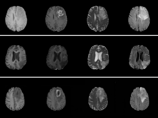
This dataset is a collection of synthetic images generated by 5 generative models (Progressive GAN, StyleGAN1, StyleGAN2, StyleGAN3, diffusion model) trained on the BraTS 2020 and 2021 datasets [1,2,3,4,5] (which share MR volumes from brain tumor patients and the corresponding tumor annotations). The trained generative models are also shared in this dataset. See our recent work [6] for more information, and a comparison of training segmentation networks with real and synthetic images.

Keywords: Radiology, Annotated, Brain, MRI, Tumor, Synthetic, Generative, GAN, Diffusion model.

Sample images

Sample images with reduced image quality. Please click to preview.

Dataset information

Short name BRGANDI
Origin Synthetic
Cite as Muhammad Usman Akbar and Anders Eklund (2023) Synthetic brain tumor images from GANs and diffusion models doi:10.23698/aida/synthetic/brgandi
Field Radiology
Organ Brain
Age span
Title Synthetic brain tumor images from GANs and diffusion models
Author Muhammad Usman Akbar
Anders Eklund
Year 2023
DOI doi:10.23698/aida/synthetic/brgandi
Status Completed
Version 1.0.0
Scans 100000 4-channel MR images per generative model
Annotations 1000000
Size 71.92GB
Resolution 1 x 1 x 1 mm
Modality MR
Scanner
Stain
Phase
References
  1. U.Baid, et al., The RSNA-ASNR-MICCAI BraTS 2021 Benchmark on Brain Tumor Segmentation and Radiogenomic Classification, arXiv:2107.02314, 2021.
  2. B. H. Menze, A. Jakab, S. Bauer, J. Kalpathy-Cramer, K. Farahani, J. Kirby, et al., The Multimodal Brain Tumor Image Segmentation Benchmark (BRATS), IEEE Transactions on Medical Imaging 34(10), 1993-2024 (2015) DOI: 10.1109/TMI.2014.2377694
  3. S. Bakas, H. Akbari, A. Sotiras, M. Bilello, M. Rozycki, J.S. Kirby, et al., Advancing The Cancer Genome Atlas glioma MRI collections with expert segmentation labels and radiomic features, Nature Scientific Data, 4:170117 (2017) DOI: 10.1038/sdata.2017.117
  4. S. Bakas, H. Akbari, A. Sotiras, M. Bilello, M. Rozycki, J. Kirby, et al., Segmentation Labels and Radiomic Features for the Pre-operative Scans of the TCGA-GBM collection, The Cancer Imaging Archive, 2017. DOI: 10.7937/K9/TCIA.2017.KLXWJJ1Q
  5. S. Bakas, H. Akbari, A. Sotiras, M. Bilello, M. Rozycki, J. Kirby, et al., Segmentation Labels and Radiomic Features for the Pre-operative Scans of the TCGA-LGG collection, The Cancer Imaging Archive, 2017. DOI: 10.7937/K9/TCIA.2017.GJQ7R0EF
  6. Usman Akbar, M., Larsson, M., Blystad, I., Eklund, A., Brain tumor segmentation using synthetic MR images - A comparison of GANs and diffusion models. Sci Data 11, 259 (2024).
Copyright Copyright 2023 Linköping University, Muhammad Usman Akbar, Linköping University, Anders Eklund
Access

Available under the following licenses, described in the License section below.

Controlled access
Free for use in legal and ethical medical diagnostics research and education.

AIDA BY license
Free for use within AIDA with attribution.

Annotation

The tumor annotation image for each 4-channel MR image was generated by each generative model.

License

Controlled access

Free for use in legal and ethical medical diagnostics research and education. Please contact the dataset provider for terms of access.

You are invited to send an access request email from your institutional account.

Clicking the access request email link above should open a draft email message in a new window, to help you provide relevant information for efficient request evaluation. If the above link does not work for you, then please click to view the suggested email text.

AIDA BY license

Copyright 2023 Linköping University, Muhammad Usman Akbar, Linköping University, Anders Eklund

Permission to use, copy, modify, and/or distribute this data within Analytic Imaging Diagnostics Arena (AIDA) for the purpose of medical diagnostics research with or without fee is hereby granted, provided that the above copyright notice and this permission notice appear in all copies, and that publications resulting from the use of this data cite the following works:

Muhammad Usman Akbar and Anders Eklund (2023) Synthetic brain tumor images from GANs and diffusion models doi:10.23698/aida/synthetic/brgandi.

U.Baid, et al., The RSNA-ASNR-MICCAI BraTS 2021 Benchmark on Brain Tumor Segmentation and Radiogenomic Classification, arXiv:2107.02314, 2021.

B. H. Menze, A. Jakab, S. Bauer, J. Kalpathy-Cramer, K. Farahani, J. Kirby, et al., The Multimodal Brain Tumor Image Segmentation Benchmark (BRATS), IEEE Transactions on Medical Imaging 34(10), 1993-2024 (2015) DOI: 10.1109/TMI.2014.2377694

S. Bakas, H. Akbari, A. Sotiras, M. Bilello, M. Rozycki, J.S. Kirby, et al., Advancing The Cancer Genome Atlas glioma MRI collections with expert segmentation labels and radiomic features, Nature Scientific Data, 4:170117 (2017) DOI: 10.1038/sdata.2017.117

S. Bakas, H. Akbari, A. Sotiras, M. Bilello, M. Rozycki, J. Kirby, et al., Segmentation Labels and Radiomic Features for the Pre-operative Scans of the TCGA-GBM collection, The Cancer Imaging Archive, 2017. DOI: 10.7937/K9/TCIA.2017.KLXWJJ1Q

S. Bakas, H. Akbari, A. Sotiras, M. Bilello, M. Rozycki, J. Kirby, et al., Segmentation Labels and Radiomic Features for the Pre-operative Scans of the TCGA-LGG collection, The Cancer Imaging Archive, 2017. DOI: 10.7937/K9/TCIA.2017.GJQ7R0EF

Usman Akbar, M., Larsson, M., Blystad, I., Eklund, A., Brain tumor segmentation using synthetic MR images - A comparison of GANs and diffusion models. Sci Data 11, 259 (2024).

THE DATA IS PROVIDED “AS IS” AND THE AUTHOR DISCLAIMS ALL WARRANTIES WITH REGARD TO THIS DATA INCLUDING ALL IMPLIED WARRANTIES OF MERCHANTABILITY AND FITNESS. IN NO EVENT SHALL THE AUTHOR BE LIABLE FOR ANY SPECIAL, DIRECT, INDIRECT, OR CONSEQUENTIAL DAMAGES OR ANY DAMAGES WHATSOEVER RESULTING FROM LOSS OF USE, DATA OR PROFITS, WHETHER IN AN ACTION OF CONTRACT, NEGLIGENCE OR OTHER TORTIOUS ACTION, ARISING OUT OF OR IN CONNECTION WITH THE USE OR CHARACTERISTICS OF THIS DATA.